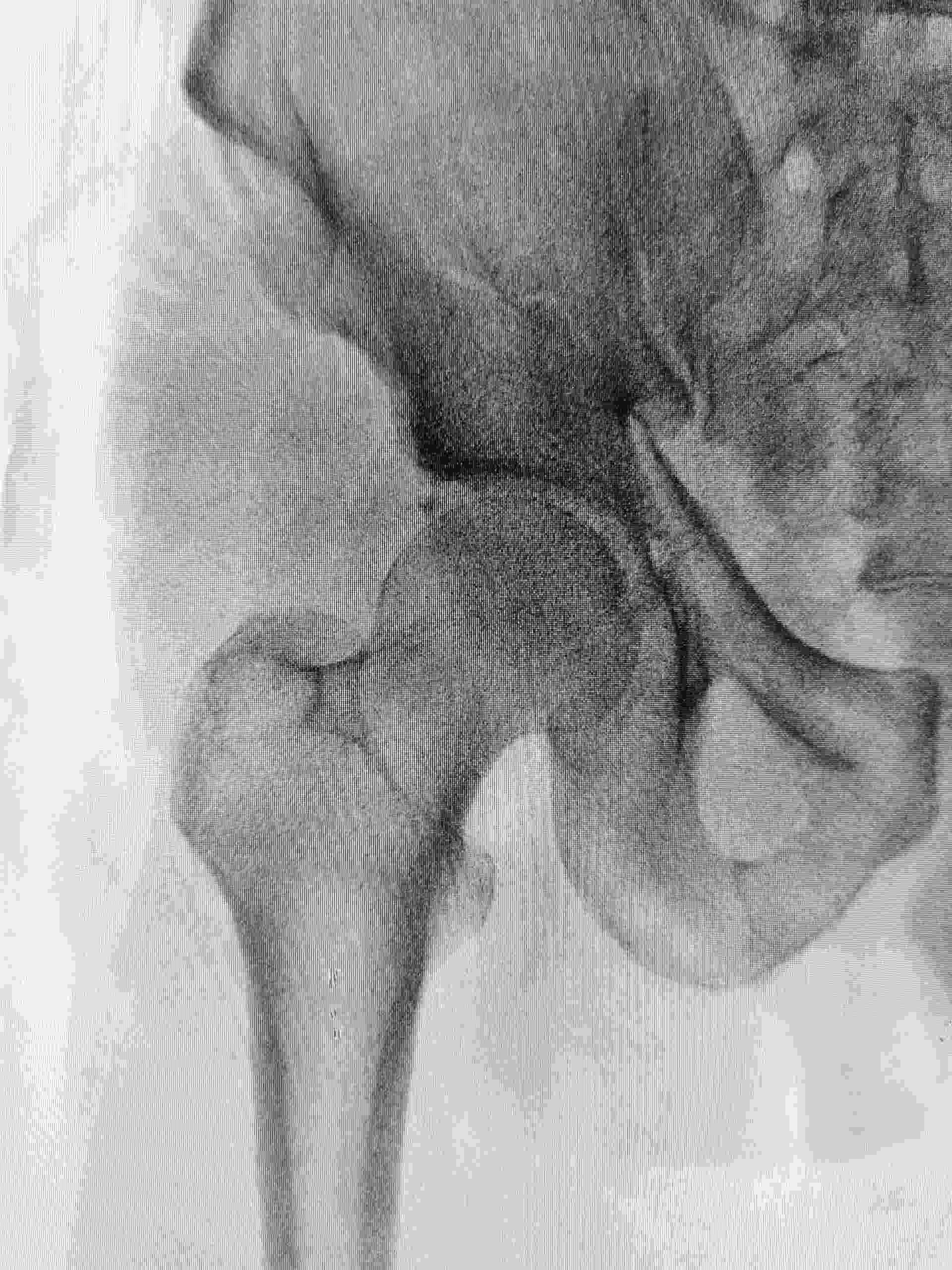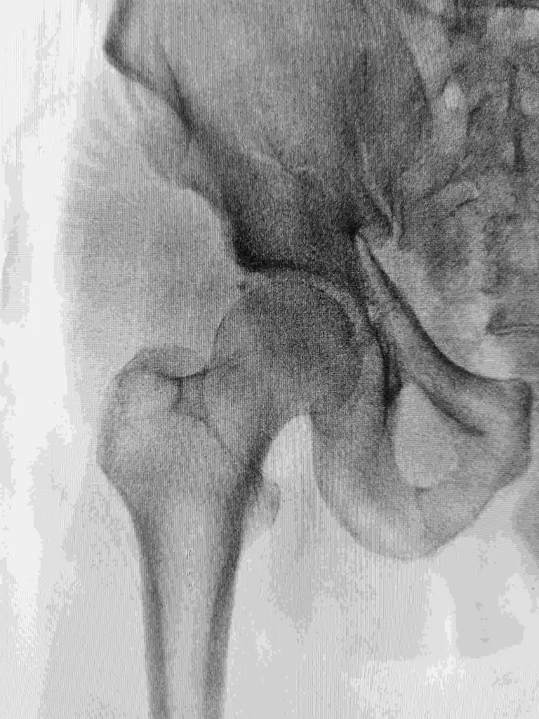

Chronic recurrent multifocal osteomyelitis is considered a form of osteomyelitis because it is present in association with a chronic state of inflammation, often associated with an underlying bone infection.
Chronic recurrent multifocal osteomyelitis is very rare.It is usually seen in the elderly or people with rheumatological disorders.t also often occurs in women, but in men is usually associated with a rheumatological disorder.However, rheumatological and osteomyelitis are sometimes seen together in a “pseudo-chondrodysplasia”.
The diagnosis is often made by finding bone infection on radiographs.
To evaluate for a more specific diagnosis, labs including X-ray and blood work can be done to identify other medical problems that may be causing inflammation.Laying down bone may also be an indicator.
Infectious disease is treated conservatively with systemic antibiotics.
Inflammation is treated with systemic steroids and surgery if required.Depending on the treatment, the course of treatment can be long, but most patients can live normal lives.Pain and radiographic changes usually disappear within a few months after treatment.Ninety to 100% of patients recover from their disease.However, about 30% of patients will experience a recurrence within a year.
If a patient is not treated for the first or second recurrence, the risk of recurrent infection is around 80%.In the United States, Chronic recurrent multifocal osteomyelitis is common and occurs more often in women than in men.Roughly two-thirds of cases of chronic recurrent multifocal osteomyelitis are seen in people over the age of 65.
Chronic recurrent multifocal osteomyelitis has been associated with autoimmune diseases, including multiple sclerosis, rheumatoid arthritis, and lupus.The normal course of chronic recurrent multifocal osteomyelitis is progressive.Progression may occur because the bone lesions weaken the bone’s ability to heal itself, become osteophytes and osteoblasts, or support infections that return.It can also be due to granulomatous, chronic vascular, fibrotic or immunosuppressive features.
Treatment is not very different from standard osteomyelitis.Most cases respond well to first line medications including antibiotics, topical steroids and oral corticosteroids.In certain cases, surgery may be required.It may cause pain, but can lead to a full recovery.
Chronic recurrent multifocal osteomyelitis is rare and is most often seen in the elderly or people with rheumatological disorders.Chronic recurrent multifocal osteomyelitis has been associated with autoimmune diseases, including multiple sclerosis, rheumatoid arthritis, and lupus.Frank and Welch (1989) reported that chronic recurrent multifocal osteomyelitis may be associated with Lyme disease and babesiosis may occur more commonly among children than adults, but the incidence of chronic recurrent multifocal osteomyelitis in children is unknown.
Although the majority of cases are in elderly patients, it can be seen in any age group.It has also been reported to be common in Caucasians.
Heme and aminopeptidase
There is also a geographical association between the causative organism and endemicity of chronic recurrent multifocal osteomyelitis.Approximately 85% of cases are associated with “Borrelia burgdorferi”, a spirochete of the genus “Borrelia”, while less than 20% are associated with “Chikungunya” virus. The bacterial causal agent can be identified through histopathological evaluation, usually found in biopsy specimens.
However, the diagnosis may also be made on the basis of serologic testing for Lyme disease.There are five serologic tests for Lyme disease, all of which have been found to have a sensitivity and specificity of 97.9%.
Another way to confirm this diagnosis is through the PCR test.If the diagnosis is in doubt, a standard fungal culture and allopurinol administration is indicated.If the cause of chronic recurrent multifocal osteomyelitis is unclear, a radiological examination with radiographic image guidance is usually helpful.
Historically, biopsy, percutaneous needle biopsy, and discography have been used to diagnose chronic recurrent multifocal osteomyelitis.Despite being a curable disease, the diagnosis of chronic recurrent multifocal osteomyelitis can be difficult due to the lack of imaging techniques and the fact that most patients do not present with symptoms in the early stages of the disease.
The first approach is to establish whether the lesion is a chronic or recurrent lesion, and in that case a specific treatment regimen would be needed.
However, there is still no diagnostic test for chronic recurrent multifocal osteomyelitis.Histopathological examination may be useful in determining the diagnosis of chronic recurrent multifocal osteomyelitis.
Positron emission tomography is the most commonly used imaging technique for evaluation.In addition, there is a limited number of studies that have been done on comparative outcome studies for chronic recurrent osteomyelitis to evaluate patient morbidity and mortality.Most patients with chronic recurrent multifocal osteomyelitis appear to improve with the appropriate treatment and the majority remain symptom-free.
Almost all patients had a complete resolution of their symptoms by the end of the initial hospitalization.A key issue in treating chronic recurrent multifocal osteomyelitis is identifying patients early in their illness, before it becomes severe.There is a high rate of relapse of chronic recurrent osteomyelitis.
Patients with chronic recurrent osteomyelitis and bone metastases have a poorer prognosis.A 2011 study found that the median survival time from the first diagnosis of chronic recurrent osteomyelitis was 17.8 years.There is significant functional disability associated with chronic recurrent multifocal osteomyelitis and the main functional limitation is loss of function.
The most common signs and symptoms of chronic recurent osteomyelitis are fever, joint pain and swelling.Symptoms may vary from day to day and it may take a while before a patient is diagnosed with chronic recurrent osteomyelitis.
Another significant symptom is joint pain or swelling, as this is usually detected more quickly.As the disease progresses patients may have joint pain associated with swelling, or even both at the same time.Other clinical findings are redness, pain or aching of joints, red patches (ughurt) on the skin, shortness of breath, swollen lymph nodes, headache, or spasms of the muscles.
Some symptoms are bilateral and some are unilateral.Additionally, skin rashes (such as erythema nodosum) and facial swelling may be present, as well as nail loss.Most of the signs and symptoms of chronic recurrent osteomyelitis can also occur during the course of the infectious period.More than half of the patients with chronic recurrent osteomyelitis presented with arthritis at the onset of the disease.
Joint pain is usually worse on the affected side.Joint pain is rarely palpable or painful, and most patients describe the joint pain as stabbing or electric-like.Less commonly, patients may have pain in the neck, lower back, shoulders, and/or upper arm.Fever is most common with chronic recurrent osteomyelitis.An elevated body temperature is associated with both secondary (temporary) infectious osteomyelitis and chronic recurrent osteomyelitis.
There is evidence that arthritis is more common in chronic recurrent osteomyelitis than in osteomyelitis caused by other infectious agents.In addition, most patients with chronic recurrent osteomyelitis experience a secondary arthritis later in the disease, which is not associated with the initial disease.Chronic recurrent osteomyelitis is usually preceded by signs or symptoms
.In chronic recurrent osteomyelitis, skin rashes (erythema nodosum) are common in the first year after the onset of the disease.
In addition, most patients with chronic recurrent osteomyelitis experience symmetrical swelling of the upper extremities at the site of the current infection.Conversely, patients with chronic recurrent osteomyelitis usually have asymmetrical swelling of the lower extremities and soft tissue deformities (i.e.; fistulas).
Secondary osteomyelitis may also occur with chronic recurrent osteomyelitis.Secondary infections are usually localized and caused by T. balneosemia.Other causes include bacterial bone abscess, dermatofibrosarcoma, non-venomous mycobacterium or other infections.
Most cases of secondary osteomyelitis occur after the primary infection has resolved.Approximately 20% of secondary infections occur in the acute phase of the disease.In most cases, there is evidence that infections are viral in nature, but are usually not recognised as such, and secondary infection is considered to be a medical pathology in its own right.
Secondary infections are associated with a number of problems; shortness of breath, headache, recurrent fever, joint pain, and sometimes skin rashes and facial swelling.Primary osteomyelitis is most commonly seen in the hips and knees
.It has been reported on the hands, legs, spine, face, abdomen and breasts.Sometimes bone changes, bone migration and the formation of bone cysts are seen.
These secondary changes are often benign.These bones can become damaged if the person who is infected is suddenly removed from their normal environment.Chronic recurring osteomyelitis is extremely rare.Since 2000, there have been only 11 confirmed cases of chronic recurrent osteomyelitis in the United States.
Chronic recurrent osteomyelitis in the United States has a relatively low mortality rate, most patients live long enough to require amputation of some of their limbs.
In one study, 13.8% of patients died while undergoing surgical or percutaneous removal of infected bone, with 4.4% of these patients dying as a result of infection in the space of 24 hours, with 6.9% dying from infection following surgery.
Another 6.9% died as a result of complications of surgery and infection, with 2.4% of these deaths being related to the causes of infection, with 4.3% from underlying conditions and 2.5% from complications of amputation.
Chronic recurrent osteomyelitis is a rare condition, usually occurring in people in their 20s and 30s.However, in some studies, it has been found to be more common in children, with 1.5–3.0% of patients developing chronic recurrent osteomyelitis by the time they are 20 years old.
It is usually found in the hip or the spine of young adults.In Australia, the incidence of chronic recurrent osteomyelitis is low, with only 15 cases reported in the five year period from 2006 to 2010.Most cases are initially misdiagnosed, as most have no accompanying signs or symptoms.
It is unclear how prevalent chronic recurrent osteomyelitis is in the US, but according to a 2010 study, it may be as high as 1.2%.Notable cases of chronic recurrent osteomyelitis in the 20th century included athletes like Florence Griffith-Joyner, Jackie Joyner-Kersee, Esther Williams and Jessica Loder Bergman.Many public figures who have died have also been diagnosed with chronic recurrent osteomyelitis, including former US president Theodore Roosevelt and former US president Richard Nixon.
Secondary osteomyelitis is more common than primary osteomyelitis.Primary osteomyelitis in children, particularly those under the age of 15, is rare.Some experts believe that this is due to the occurrence of pressure sores and infection in the space around the buttocks which are more likely to become infected during childhood.
Around 1 in 40 to 1 in 60 adolescents will develop osteomyelitis at some point in their life.Some cases of primary osteomyelitis in children can be very serious, involving the amputation of a leg or fingers, or even death.Secondary osteomyelitis in children is typically more severe.In this type of osteomyelitis, the bone abscess develops due to an infection occurring outside of the original location of bone infection.
Secondary osteomyelitis in children can present as primary infection around a secondary site.It can also be due to primary infection of a secondary site.Most cases of secondary osteomyelitis in children are due to bacterial infections.Secondary osteomyelitis in children is more likely than in adults to develop complications.
This can include a reaction to antibiotics, sepsis, or septic arthritis.The serious complications that can arise are much more common in children than in adults, and can lead to death or permanent disability.Other serious complications that have been reported in children are liver failure, kidney failure, and bone lesions, especially at the site of the bone infection.Chronic recurrent osteomyelitis in adults is rare.
The National Institutes of Health (NIH) estimates that less than one case is diagnosed each year in the US.It is most commonly seen in young adults, with the age of incidence of around 20 years.As of 2011, the incidence of chronic recurrent osteomyelitis was estimated to be 0.21 cases per 100,000 adults per year.In the United States, the National Osteoporosis Foundation estimates the incidence of osteomyelitis to be 6 per 100,000 people per year.
Worldwide, chronic recurrent osteomyelitis is much less common.One estimate based on data from 2010 suggests the prevalence to be less than 1 case per 100,000 people per year.The incidence in Australia is estimated to be 1 case per 10,000.
The most common site of infection in people with chronic recurrent osteomyelitis is the buttocks.More than half of all cases of chronic recurrent osteomyelitis are as a result of eroding bone, most commonly occurring in the back.A small number of cases are caused by rickets, but this is uncommon.
Over 80% of cases have a definitive cause, with the most common being osteomyelitis due to sexually transmitted disease or bacterial infection.Chronic recurrent osteomyelitis is not caused by the autoimmune disease known as rheumatoid arthritis.Researchers still do not know the exact cause of chronic recurrent osteomyelitis, and therefore, there is no treatment that is specific to this condition.
There are a number of treatment options for chronic recurrent osteomyelitis.
The main treatment option is surgery.There are many different surgeries available depending on the location of the infection and other factors.Permanent debridement is performed to remove the infection, or bone necrosis caused by infection.Typically, the infection surrounds a nerve, such as that of the genital area or the breast.
The infection is covered with flesh, usually removed during surgery.Arthroplasty is a minimally invasive surgery used to replace the fractured femur.While this is done, the previously healthy bone around the femur is harvested and used to replace the affected bone.
A growing number of cases of chronic recurrent osteomyelitis in the rectum occur due to Erythema nodosum, a bacterial infection.
While this is not an infection of the bone itself, it can be treated similarly.


